

Most skin cysts are harmless sacs filled with keratin rather than anything dangerous. They do not clear on their own, and draining one without removing its lining lets it refill. Cysts are evaluated and removed at Coastal Dermatology + Aesthetics in San Luis Obispo.
A cyst is a sac, and the sac is the part that matters
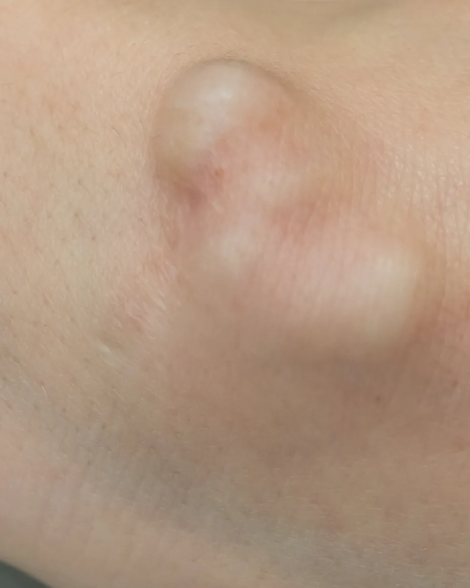
A skin cyst is a closed sac under the skin. [9] Most of the ones people bring in are epidermoid cysts, which are benign, sit just under the surface, and are filled with keratin, the same protein your skin and hair are made of. [1] They form from the upper part of a hair follicle. [1] Often there is a small dark pinpoint opening in the center of the lump, which is the follicle it came from. [9] [10]
The word “sebaceous” attached to these is a misnomer, and it is worth clearing up because it is on most of the pages you will read. These cysts do not involve the sebaceous gland and they do not contain sebum. [1] The contents are keratin and cellular debris, which is where the thick, pale, strong-smelling material comes from when one ruptures. [1] [10]
The second common type is the pilar cyst, also called a trichilemmal cyst, which comes from the outer sheath of the hair root and contains keratin. [3] They mostly affect the skin of the scalp, though they can also appear on the face, neck, arms and legs. [3] [8] They present as flesh-colored, smooth, mobile, firm, well-circumscribed nodules, most commonly as multiple lesions, and they may be inherited as a dominant trait, so more than one person in a family often has them. [3] [8] They do not give rise to malignant lesions. [3]
What a cyst usually looks and feels like [9] [10]
- A round lump under the skin that moves when you press it
- Most often on the face, neck, trunk or scalp
- Frequently a tiny pinpoint opening in the center
- Slow growing and usually painless
- Thick, pale, strong-smelling contents if it opens
- Red, warm, tender and swollen if it has ruptured or become inflamed
The reason a cyst comes back. Draining the contents without removing the lining leaves the structure that makes them, so it refills. [2] [6] [7] Taking the sac out intact is what lowers the chance of that. [1] [3]
Where cysts come from
The follicle closes over
Epidermoid cysts form when surface skin cells end up underneath the skin surface instead of shedding from the top. [9] Those cells keep producing keratin with nowhere to go, and the sac fills from the inside. [10] The cause is often simply unknown. [9]
Injury, irritation and inheritance
Skin injury or irritation of a follicle can start one. [10] Pilar cysts have a different story: they may be inherited as a dominant trait, [3] and a tendency to develop them can be passed down, which is why people who get one often have several and often have a parent who had them too. [8]
Rupture, and what it feels like
When a cyst ruptures, keratin is displaced into the dermis and the surrounding tissue and the body reacts to it. [1] [3] A ruptured cyst can closely resemble a boil, with tenderness, redness and swelling. [1] An inflamed cyst is usually settled down first and removed afterward, because removal goes better once the area is quiet. [1]
Where pilar cysts mostly appear. [3] They are firm, smooth and mobile, they most commonly turn up as multiple lesions, and they may be inherited. [3] [8]
Why a cyst that was drained comes back
This is the single most useful thing on this page, and it is the reason people end up having the same lump treated two and three times.
| What was done | What happens next |
|---|---|
| Nothing | Some shrink. Others keep growing. Without treatment a cyst can stay for the rest of your life [6] |
| Squeezed at home | The contents move rather than leave, and the risk is infection and a lump that refills [6] |
| Drained or lanced only | Fast relief from pressure, and the lining stays behind, so it can fill again [6] [7] |
| Removed whole, sac included | Removing the entire lining is what lowers the chance of it returning [1] [2] [3] [9] |
A cyst is a sac with contents, and the two are not the same problem. Emptying it addresses the contents. The sac is a functioning structure that keeps producing keratin, so as long as it is in place the lump has a way back. [6] [7] Incomplete excision is the main reason a cyst reappears in clinical practice. [2]
What removal actually involves. The area is numbed with a local anesthetic, so you are awake and the procedure is not painful. [6] A small incision is made, sized to the cyst, the cyst is lifted out in one piece with its lining, and the skin is closed with sutures. [7] Small sites heal in a few days to a couple of weeks. Larger ones with a longer incision can take several weeks. [7] Any incision leaves a mark, and where it is placed and how it is closed are part of the plan rather than an afterthought. [10]
Where a cyst is actively inflamed or infected, the sequence changes. Complete excision is delayed while an active infection is present, and an initial drainage or an injection to reduce swelling comes first, with full removal scheduled once the inflammation has resolved. [1] [10] That is standard practice, not a delay.
The kinds we see, and where they show up
| Type | Where it usually appears | Worth knowing |
|---|---|---|
| Epidermoid cyst | Face, neck and trunk [10] | Keratin filled, from a hair follicle, often with a pinpoint center [1] [9] |
| Pilar, or trichilemmal, cyst | Mostly the scalp, and sometimes the face, neck, arms and legs [3] [8] | Firm and mobile, most commonly multiple, and possibly inherited [3] [8] |
| Digital mucous cyst | Over the last joint of a finger or toe, near the nail [5] | Originates from the joint, is often associated with arthritis in that joint, and can cause a deep lengthwise groove in the nail [5] |
| Acne cysts and nodules | Face, chest and back | A different thing entirely. These are inflammatory acne cysts and nodules, not sacs, and they are treated as acne |
The last row causes real confusion, and it is worth separating. A deep, painful acne lesion is sometimes called a cyst, and it is not the kind of cyst this page is about. Those are handled as part of acne treatment. The lumps on this page are sacs with linings, and removing the lining is the whole point.
The digital mucous cyst is the one with a knack for coming back, and the reason is the same as everywhere else on this page: it grows from a stalk connected to the joint, and removing the cyst along with that stalk is what settles it. [5] Where the stalk is left, it returns.
Services are prompt, friendly, professional and most of all comfortable…it was an extraordinary experience.
The Coastal difference
Not everything that feels like a cyst is one
Most lumps under the skin are exactly what they look like. A few are not. The list of things a suspected cyst can turn out to be is longer than people expect, and it includes lipomas, neuromas, other benign growths, and skin cancers. [2] A lump that moves, that feels familiar under a fingertip and that has been there a while is usually ordinary. Sorting the usual from the unusual is a five-minute job for someone who does it every week.
That is the argument for having a lump looked at by a dermatologist rather than removed by whoever is nearest. When a cyst is taken out here, the tissue is examined under a microscope, because while the great majority of these cysts are benign it matters to send the excised sample for evaluation and confirm there is no malignancy. [1] The lining and the contents are exactly what a pathologist reads to classify it. [4] Most of the time that confirms the obvious. Occasionally it identifies a lump that turns out to be something else, and identifying it early is the entire value of doing it that way.
What happens to a cyst after it is removed here. The lining and the contents are what classify it under a microscope. [4] Most results are unremarkable, which is the point of checking. [1]
When to see a dermatologist
Book an appointment if a lump: [10]
- Is growing quickly, or several have appeared
- Has opened, or is red, warm, swollen and tender
- Is painful, or shows signs of infection
- Sits somewhere that keeps getting caught, scratched or knocked
- Is somewhere unusual, such as a finger or toe
- Bothers you because of how it looks
Signs a cyst has become infected [9]
- Redness of the skin over it
- Tenderness or soreness
- Warmth in the area
- Grayish white, cheesy, foul smelling material draining from it
Contact us about any new growth you notice. [9] In most cases the diagnosis is made by examining the skin. [9] Most cysts are not dangerous and do not need to be treated unless they cause symptoms or show signs of inflammation, [9] so an appointment is often about knowing what something is and deciding what you want to do about it.
What helps, and what to leave alone
Worth doing
- Leave it alone. Do not try to pop or drain a cyst yourself, since that risks infection and it will likely regrow [6] [10]
- A warm, moist cloth held over the area can help a cyst drain and heal [10]
- If a lump becomes red, warm and tender, have it seen rather than waiting it out [10]
- Bring in a lump that keeps getting caught by a collar, a razor or a hairbrush before it is inflamed, since removal is more straightforward when the area is quiet [1]
After removal
- Keep the site clean and covered as directed, and follow the suture care instructions you are given
- Small sites heal in a few days to a couple of weeks, and larger ones can take several weeks [7]
- Any excision leaves a mark, and that trade is discussed before anything is scheduled [10]
- New cysts can appear elsewhere, particularly with pilar cysts, which most commonly come in numbers [3] [8]
Worth knowing
- Most skin cysts are benign [1] [3]
- The contents are keratin, not sebum [1]
- A cyst that is emptied but not removed has a route back [6] [7]
- Pilar cysts may be inherited, and one is often followed by others [3] [8]
- A digital mucous cyst near a nail settles when it is removed along with the stalk connecting it to the joint [5]
Treatments we offer
Evaluation, in-office removal, and where a lump needs an answer rather than a procedure, the test that gives one. Pick a treatment to see how it works.
What treatment costs
Evaluation and removal of a skin cyst are medical care rather than a cosmetic service, and are handled differently from the aesthetic treatments on this site. Our office will go through what applies to you when you book.
Where tissue is sent for examination, laboratory fees are billed separately by the laboratory, and we will tell you at the time that anything is being sent.
What is actually inside
Keratin, a skin protein, not sebum, which is why “sebaceous cyst” is a misnomer [1]
The most common two
Epidermoid cysts, which arise from a hair follicle, and pilar cysts, which are usually on the scalp [1] [3]
Why one comes back
The lining was left behind. Removing the sac intact is what lowers the chance of it returning [1] [2] [3]
Whether it will resolve on its own
Some shrink and others keep growing. Without treatment a cyst can stay for the rest of your life [6]
Cyst removal frequently asked questions
How long a cyst can stay without treatment. [6] Some shrink on their own and others keep growing, and neither one resolves the sac.
Offered at
Have a lump you want identified? Request an appointment
Fill out the form below, book online in Santa Maria (opens online scheduler), or give us a call. Cosmetic and aesthetic appointments need no referral. For medical dermatology, we are accepting new patients by referral from your primary care physician; send the referral across and then use the same form.
Sources and medical review
1. StatPearls, National Center for Biotechnology Information. “Epidermoid Cyst.” States that an “epidermoid cyst, also known as a sebaceous cyst, is a benign encapsulated, subepidermal nodule filled with keratin material,” that “epidermoid cysts are derived from the follicular infundibulum,” that “the term sebaceous cyst is commonly used; however, it is a misnomer, as it does not involve the sebaceous gland,” and that “cysts may progress slowly and remain present for years.” On rupture, states that “when the cysts rupture, an inflammatory reaction occurs due to the displacement of soft, yellow keratin into the dermis and surrounding tissue,” and that “epidermoid cysts are usually asymptomatic; however, if ruptured, they may closely resemble a furuncle with tenderness to palpation, erythema, and swelling.” On treatment, states that “the most effective treatment involves complete surgical excision of the cyst with the cyst wall intact,” that “removing the entire cystic lining is important in decreasing recurrence,” and that “the complete excision should be delayed if an active infection is present,” with an initial incision and drainage indicated in those cases. On pathology, states that “while the majority of these cysts are benign, it is important to send the excised sample for evaluation to ensure that there is no malignancy,” and that “histological examination reveals an epithelial-lined cyst filled with laminated keratin located within the dermis.” https://www.ncbi.nlm.nih.gov/books/NBK499974/ · Medical Reference
2. StatPearls, National Center for Biotechnology Information. “Epidermal Inclusion Cyst.” States that the differential diagnosis “includes pilar cyst, lipoma, abscess, neuroma, benign growths, skin carcinomas, metastatic cutaneous lesions, pilomatrixoma, ganglion cyst, neurofibroma, dermoid cyst, brachial cleft cyst, pilonidal cyst, calcinosis cutis, pachyonychia congenita, steatocystoma simplex, and steatocystoma multiplex,” and separately that it “also include lipomas, neuromas, benign cutaneous growths, metastatic skin lesions, squamous cell carcinoma of the skin, basal cell carcinoma of the skin.” States that “epidermoid inclusion cysts can be confirmed by histologic examination,” that “there is a risk of the cyst reoccurring if the capsule is not completely excised during the surgical procedure,” and that “the main complication seen in clinical practice is reoccurrence due to incomplete excision.” https://www.ncbi.nlm.nih.gov/books/NBK532310/ · Medical Reference
3. StatPearls, National Center for Biotechnology Information. “Pilar Cyst.” States that “pilar or trichilemmal cysts are common dermal cysts,” that “the cysts contain keratin and are outlined by stratified squamous epithelium” similar to the outer root sheath of the hair follicle, that “they mostly affect the skin of the scalp,” that they “usually present as flesh-colored, smooth, mobile, firm, and well-circumscribed nodules,” that “most commonly, they are multiple lesions,” that “trichilemmal cysts may be inherited as an autosomal dominant trait,” and that “trichilemmal cysts never give rise to malignant lesions.” States that ruptured cysts leak contents into the dermis “leading to the formation of a foreign-body reaction,” that “the mainstay of treatment is radical surgical excision of the lesion, including the wall of the cyst,” and that “if the surgeon does not remove the sac completely, then the recurrence rate will be high.” https://www.ncbi.nlm.nih.gov/books/NBK534209/ · Medical Reference
4. StatPearls, National Center for Biotechnology Information. “Dermatopathology Evaluation of Cysts.” States that “a structured approach to the histopathologic diagnosis of cutaneous cysts involves the assessment of the following features: Epithelial lining, Cyst contents, Ancillary histopathologic features,” and that pilar cysts “usually present as a solitary nodule most commonly located on the scalp of a female adult.” https://www.ncbi.nlm.nih.gov/books/NBK603737/ · Medical Reference
5. StatPearls, National Center for Biotechnology Information. “Digital Mucous Cyst.” States that “digital mucous cysts (also known as digital myxoid cysts or digital mucous pseudocysts) are ganglion cysts that originate from the dorsal aspect of the distal interphalangeal joint (DIP),” that they “typically manifest as slow-growing masses located in the subcutaneous tissue overlying the dorsal aspect of the DIP joint,” that they “most commonly affect the fingers but can also affect the toes and are frequently associated with underlying DIP joint osteoarthritis, although arthritis may be absent in some cases,” and that “cysts in this location cause deep longitudinal grooves of the nail plate.” On recurrence, states that “after surgical excision of digital mucous cysts, recurrence occurs in approximately 2%, provided the excision includes the stalk of the cyst,” and that removal without the stalk carries a substantially higher recurrence rate. https://www.ncbi.nlm.nih.gov/books/NBK559092/ · Medical Reference
6. Cleveland Clinic. “Sebaceous Cysts (Epidermal Inclusion Cysts).” States that “an epidermal inclusion cyst (epidermoid cyst) is a fluid-filled pocket under the surface of your skin,” that the term sebaceous cyst is misleading because keratin and cell debris rather than sebum fill these cysts, and that “some cysts decrease in size, while others continue to grow until you get treatment. Without treatment, you may have the cyst for the rest of your life.” Advises: “don’t try popping or draining the cyst yourself. This could cause an infection, and the cyst will likely grow back (recur).” States that drainage “won’t resolve the cyst, since your provider won’t remove the cyst capsule,” and that “the removal of the capsule prevents the cyst from growing back.” Notes that removal uses “a local anesthetic (you won’t be asleep and you won’t feel pain).” https://my.clevelandclinic.org/health/diseases/14165-sebaceous-cysts · Medical Organization
7. Cleveland Clinic. “Cyst Removal.” States that “your provider removes the entire cyst from under your skin, including the lining (sac),” that “this lowers the chance of it coming back,” and describes the steps as making “a cut in your skin based on the size of the cyst,” removing “the entire cyst in one piece,” and closing “the cut in your skin with stitches.” States that “some cysts can come back, especially after drainage. This happens when the sac or lining isn’t fully removed,” and that “small cysts that don’t need stitches usually heal in a few days to a couple of weeks. Larger cysts with bigger cuts may take several weeks or even months.” https://my.clevelandclinic.org/health/procedures/cyst-removal · Medical Organization
8. Cleveland Clinic. “Pilar (Trichilemmal) Cyst.” States that “pilar cysts are relatively rare noncancerous (benign) cysts that grow from your hair follicles,” that “most pilar cysts surface on your scalp (head), but they can appear on your face, neck, arms and legs,” that “for the most part, trichilemmal cysts are benign, meaning they don’t become cancerous,” that “pilar cysts are hereditary, meaning you may inherit a tendency to develop these cysts,” that “if you have one cyst, more may develop,” and that “you may end up with an infection if you try to pop or slice open your cysts.” Notes that a provider may “examine the cysts under a microscope to confirm your cysts aren’t cancerous.” https://my.clevelandclinic.org/health/diseases/23092-pilar-trichilemmal-cyst · Medical Organization
9. MedlinePlus, U.S. National Library of Medicine. “Epidermoid cyst.” States that “an epidermoid cyst is a closed sac under the skin, or a skin lump, filled with dead skin cells,” that they “are very common” and their cause “is unknown,” that they form “when elements of the surface skin get under the skin surface,” that “the main symptom is usually a small, non-painful lump beneath the skin,” and that “it will often have a tiny hole or pit in the center.” Lists infection signs as “skin redness,” “tender or sore skin,” “warm skin in the affected area,” and “grayish-white, cheesy, foul-smelling material that drains from the cyst.” States that “epidermoid cysts are not dangerous and do not need to be treated unless they cause symptoms or show signs of inflammation,” that “in most cases, your health care provider can make a diagnosis by examining your skin,” that “they may return if they are not completely removed by surgery,” and to “contact your provider if you notice any new growths in your body.” https://medlineplus.gov/ency/article/000842.htm · Government Organization
10. Mayo Clinic. “Epidermoid cysts.” States that “epidermoid cysts are harmless small bumps beneath the skin,” that “epidermal cells form the walls of the cyst and then secrete the protein keratin into it,” that “keratin is the thick, cheesy substance that can leak from the cyst,” and that “sometimes this type of cyst forms due to irritation or injury of the skin or a hair follicle.” Lists signs including “a small, round bump under the skin, often on the face, neck or trunk,” “a tiny blackhead plugging the central opening of the cyst,” “a thick, smelly, cheesy substance that leaks from the cyst,” and “an inflamed or infected bump.” Advises seeing a health professional for a cyst that “grows or multiplies rapidly,” “breaks open,” “is painful or infected,” “is in a spot that keeps getting scratched or bumped,” “bothers you because of how it looks,” or “is in an unusual spot, such as a finger or toe.” Recommends “placing a warm, moist cloth over the area to help the cyst drain and heal” and “not squeezing a cyst yourself.” States of drainage that “cysts might recur after this treatment,” and of excision that “your healthcare professional removes the entire cyst,” which “is safe and effective and often prevents the cyst from regrowing,” but “may leave a scar.” Also notes that “injecting a steroid into the cyst can ease swelling and inflammation.” https://www.mayoclinic.org/diseases-conditions/epidermoid-cysts/symptoms-causes/syc-20352701 · Medical Organization
Content on this page is for educational purposes and is not a substitute for professional medical advice, diagnosis or treatment. Most skin cysts are benign, and not every lump under the skin is a cyst. Removal is a minor surgical procedure that carries the usual risks of any incision, including bleeding, infection and scarring, all of which are discussed before treatment. Cysts can recur, particularly where a cyst has ruptured before removal or where the lining cannot be taken out intact. Tissue removed here is examined by a laboratory. Individual results may vary.